by BARRACUDA
Before It’s News
July 9, 2012
Revelations From A Man Who Helped Design Morgellons Disease
There are two videos at Carnicom.com about Morgellons Disease. These are Important videos and must be watched.
The first video:
“Introductory Remarks”
http://www.carnicom.com/morgvid1.htm
The 2nd video:
“Morgellons 2nd Session”
http://www.carnicom.com/morgvid2.htm
These 2 videos are well done and well researched by Clifford Carnicom and Dr. Gwen Scott. The information in these videos affects every individual in this country. They are perhaps the two of the most important videos ever to be made. If you have not viewed these videos you need to do it ASAP. These videos are not casual entertainment they are about all of us. Not just a few people with a weird disease called Morgellons, but about all of us. This information personally affects every person you love and care about. It is about your grandchildren, your family pet, your loved ones and even people down the street that you don’t care about. We are all being subjected to this. It is in the very air we are breathing, and the food we are eating. That is certain. There are more vectors beyond even that. To put it in simple terms:Continued below.
We May All Have Morgellons Disease and It Is Man Made!
Many people viewing these videos may have overlooked one of the most import pieces of information that was included in each. Someone has confessed to participation in at least one aspect of the formulation of this disease. It is not to be overlooked or tossed aside. I do believe that Dr. Gwen Scott has been contacted by a credible source who has admitted his part in creating Morgellons Disease. There is still so much conjecture as to the origins of Morgellons that perhaps Gwen’s statements will impact someone who is still pretending that this disease in not a deliberate assault on all humankind. It is important to make sure everyone will hear or read this message. For this reason I have transcribed the relevant points she made about the origin of Morgellons disease from her videos with Clifford Carnicom. Perhaps seeing it all in print will give it more credibility. Since it is the spoken word and not text originally I wrote it verbatim as she spoke it without any changes.
I give great credit to Dr. Gwen Scott for putting forth this vital information and for her selfless work for she has done for the Morgellons community. She is herself a sufferer of the extreme symptoms of the disease. Likewise, I am proud to know Clifford Carnicom and count him among the true heroes of our time for his selfless dedication to this research and getting the truth out there in spite of the personal dangers that can come with being an honest man in these dark times. Clifford has revealed the biggest “dirty little secret” known to man.
________________________________________________________
Here is the written transcript from a small portion of the spoken dialogue by Dr. Gwen Scott from the above “Introductory Remarks” video regarding who responsible for origins of Morgellons disease.
http://www.carnicom.com/morgvid1.htm
“Introductory Remarks”
Dr. Gwen Scott States:
“I began to see there are a lot of components. It is a very compound complex network of pathogens. Organic, inorganic and all kinds of things.
We have fibers, heavy metals, bacteria, funguses, viruses that seem to be working somehow synergistically. None of them good to the Human.”
“I was able to touch base with one of the people who actively was involved in the design of some of this and I think at the time he thought he was doing this country some good – and in retrospect now realizes, perhaps not. Now he’s trying very hard to help anybody who’s conscious of what’s going on. He did tell me that most of these pathogens have been genetically altered so that your immune system doesn’t know that they’re there. They’re cloaked, they’re different, they can overcome. I won’t go into the specifics but just let’s say these are not your average bacteria, virus, fungus – and of course heavy metals are not average in the human body anyway – and these fibers, these unusual fibers are wires that we’re seeing. People have said, “Well, where’s it coming from?” Clearly, there have been enough samples to know and Mr. Carnicom, Clifford, just made that connection for us through the air. Some of them are coming to us through the air, with deliberation through the air supply. I have been told by this same gentleman it is also being delivered to us by our food supply. Particularly commercial preprocessed food”
Gwen also went on to say she believes that another vector of transmission is through vaccines.
Here is the written transcript from a small portion of the dialogue by Dr. Gwen Scott from the above “2nd Video” regarding her contact with one of the designers of Morgellons Disease.
http://www.carnicom.com/morgvid2.htm
“Morgellons 2nd Session”
Dr. Gwen Scott states:
“Since our last discussion I did have a gentleman who was involved in some of this (Morgellons design) call me. When he was involved he felt he was doing something to help the soldiers in the field in this country. He was told these things would be sprayed, aerosol sprayed, from planes on the enemy and they would save soldier’s lives – and then it occurred to him when he became aware and began to see Clifford’s work and other work that, oops,
maybe that’s not the case. So now he’s trying really hard to help out anybody that he can find that’s trying to do the work. -and he explained something to me that I knew that I had kind of forgotten. Every organ in your body has a specific frequency and it operates at that frequency. -and when you interrupt that frequency electromagnetically, you can create all kinds of serious (even unto death) problems.
He also talked about areas of the brain and mind control. -and as Orwellian as that may seem, apparently, scientifically it is very real. We know from Clifford’s work, for years now, the electromagnetic properties of what’s happening in our air supply as a result of the manipulation and the manipulation of that. Beyond that he talked about then confirmed to me the heavy metals, the barium, the titanium, the aluminum. None of which, trust me, are good for the human. He talked about the biological. He said all of it had been altered. Some of it (and Clifford had eluded to earlier in the video) can kind of escape your immune system, cloak itself in one form or another. Again (he spoke of) the electromagnetics and also the lack of oxygen, the displacement of oxygen. The more you displace oxygen out of the air supply with particulates, (it could be Cornflakes, it doesn’t matter) the mortality rates go up concurrently. We are operating on a very low level of oxygen.”
Dr Gwen Scott who is also a Morgellons sufferer shares her excellent holistic protocol for Morgellons and a Flu Protocol with others at the following link. ArizonaSkywatch
While you are at Arizona Skywatch check out the Morgellons photos and research from my friend Blue “HERE” .
Source Morgellonsexposed.com
August 21, 2012 | Categories: chemtrails, food, geo-engineering, health, morgellons, news, tyranny | Leave a comment
by Kathryn A. Augustyn
Morgellons Research Group
August 17, 2012
The ‘Brown Rain’ Event
Remember the odd “rain” that left Chico’s cars streaked with brown ash-like residue? What follows is MRG’s report from Marla Crites (and Betty Credit):
The Weather Event of May 14, 2012
In the words of one of our members who was awake during the night of the strange rain…
“It was called rain, however, myself, my son, and others I have spoken to, did not feel the wetness of the drops. The water seemed to dissipate at touch. At 2:30 a.m., my son woke me up to tell me of the powder that had covered my car. At the time, I thought it was pollen from the trees, blown off from the rain. However, upon morning, I definitely had questions about what had actually happened the night prior. My car, and all others I saw in my immediate area, were covered with a tan-colored light ash. It was such a fine powder; yet, I could not drive my car without washing my window. One of the creepiest experiences was driving to the store. Every car I passed, that had obviously been exposed was covered with the same thing. Then, to top things off, I had to go to the car wash to clean my car, because it looked like it was covered in volcanic ash!! When I got to the car wash, there was a line of cars that was unprecedented. Every car was covered with the same thing. By the time I exited the wash, the cars in line were uncountable. No explanation, no one seemed bothered by this ‘EVENT’.”
Adding to her description: Each car was not only covered in a fine tan powder, but there were often streaks through the powder as if made by water. The “ash” was not easy to scrape off the windshields, but I managed to obtain several samples for testing.
The Official Explanation
The Enterprise Record newspaper ran a story the next day in which Anthony Watts, longtime local meteorologist, was interviewed. He surmised that agricultural activity had raised a great deal of dust which an updraft carried to the upper atmosphere from where it fell as the “brown rain”. It is true that plumes of dust did rise as farmers worked the dry fields. (although this happens every year) I enquired of the Air Quality Board and the Public Health Environmental people as to what they thought had caused this strange event. They all lined up behind Anthony’s explanation and had no plans to test any of the resulting powder.
Chico Sky Watch Tests
A ranch manager in Durham tasted (yes!) the powder, said it was vile and not dirt or pollen. A CSW member poured purified water on her solar panel, caught it at the bottom in a clean bowl, and took it to Basic Lab. Results were 52,000 ppb aluminum, 480 barium and 413 strontium. To put these numbers in perspective, aluminum should be 0.5 ppb and 1,000 ppb is the EPA Maximum Contaminant Level and Mandatory Action Level. There should be no barium or strontium in rain. So these numbers are exceedingly and alarmingly high.
We sent a sample of the powder to Larry Meyer in Oregon who cultured it and sent the growths on to Pro-Lab in Florida. Results were that 6 different, not uncommon molds were present. The molds are known to cause asthma, hay fever and allergies. Links to both lab reports follow.
Basic Lab Report
http://www.chicoskywatch.org/assets/tests/basic-lab-report-6-22-12-1a079e347fadc0f6c3b23de1e6395cf8.pdf
http://www.chicoskywatch.org/assets/tests/pro-labs-report-6-6-12-1fca340ea69f24ad46fbfc27f6a5a9e0.pdf
Unanswered Questions:
1. Is there any way to distinguish natural soil based aluminum from the aluminum oxide that is a component of aerosol spray?
2. How likely is it that dust from farming activities would account for the 52K ppb in the rain?
3. How abnormal for rainfall is the type and amount of molds identified by Pro-Lab?
4. Is there mold in “normal” rainwater?
See http://www.chicoskywatch.org/tests for more information.
Chico Rain Lab Report
(Gratitude to Larry Meyer, MRG Senior Research Associate)
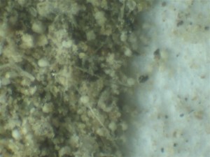
Recently, Morgellons Research Group (MRG) received an interesting specimen from Chico Skywatch, collected from a most unusual rainstorm which fell over a sizable area of Butte County, California. Marla Crites of Chico Skywatch in submitting the specimen wrote that “brown rain” had fallen on this locale on May 14, 2012.
Chico Sample As Received by L. Meyer
Chico sample as it began to bloom in agar (L. Meyer)

The Chico sample as sent to Pro-Labs (L. Meyer)

MRG performed a microscopic examination/photography of the brown finely granular sample and placed a portion in agar to observe what might grow. The resulting “bloom” was sent to our laboratory for identification. Six varieties of fungal material were identified.
The ProLab Report
Prolab report on chico rain
Dr. Richie Shoemaker, MD of Pocomoke, Maryland, in his book MOLD WARRIORS, states that there is evidence several fungal varieties that may have been “weaponized”.
At this time, Morgellons Research Group wishes to initiate a research project to study rain and snow precipitates and is pleased to invite you to precipitate. It is our desire to collect data from the various areas where Skywatch groups are active. Data obtained would be shared with participating groups and published on MRG’s website.
The Second Collection Sent to the Lab
by Larry Meyer
On June 28th, I used a clean petri dish to capture the first five raindrops to fall that day at my location on the Oregon Coast. Agar was added and a bloom began within a day. Shown is one of several varieties of Fungus from that rain:


THE FINDINGS
ProLab Report sent to L. Meyer
2nd prolab report first 5 drops of rain
In July 2012, Larry Meyer also collected this specimen from rain. An artifact found in rain on the Oregon Coast- July 3, 2012 – 0.5 x 0.5mm.

Image Credit to Larry Meyer
Six (6) species of Fungi and One (1) yeast were found in both of these samples:
Cladophialophora was found in Portland, Oregon

For comparison, one can see that a mixture of these could be devastating, not only to crops, ocean life but human life as well. How did these spores and mycelia come together and form a brown rain? Yellow rain is very similar. Red rain is from an algae.
I. Cladosporium, (found in both Samples of Brown Rain) Examples of Cladosporium
A . Cladosporium …..C Herbarum

Allergen Exposure
Spores of Cladosporium spp. probably occur more abundantly worldwide than any other spore type and are the dominant airborne spores in many areas, especially in temperate climates. (1, 2, 3, 4) Although C. cladosporioides may be the most prevalent airborne species, C. herbarum frequently dominates indoor and outdoor air and is a major source of fungal inhalant allergens. (3, 5)There are about 500 species of Cladosporium. Many are saprophytic on plant litter.
C. herbarum is widely distributed in our environment and is a major source of fungal inhalant allergen. (4) C. herbarum is one of the most common environmental fungi to be isolated worldwide. It occurs abundantly on fading or dead leaves of herbaceous and woody plants, as a secondary invader on necrotic leaf spots, and has frequently been isolated from air, foodstuffs, paints, textiles, humans and numerous other substrates. It is also known to occur on old carpophores of mushrooms and other fungi and as a common endophyte, especially in temperate regions. Under favourable climatic conditions C. herbarum also germinates and grows as an epiphyte on the surface of green, healthy leaves. (6)
http://www.phadia.com/en/Allergen-information/ImmunoCAP-Allergens/Molds-and-other-Microorganisms/Allergens/Cladosporium-herbarum-/
**************************
B. Cladosporium: C. carrionii and C. yegresii

Fig. 7. Conidial morphology in selected branches of (upper row: A-C) C. carrionii, strain CBS 260.83; (lower row: D-F) C. yegresii, strain CBS 114405. In this respect the two species are identical. Scale bar = 10
http://www.biomedsearch.com/nih/Molecular-analysis-pathogenicity-Cladophialophora-carrionii/18491001.html
C. Cladosporium elatum, Cladosporium herbarum, Cladosporium sphaerospermum, and Cladosporium cladosporioides.


Cladosporium is the genera most frequently encountered in both outdoor and indoor air. It is frequently found in elevated levels in water-damaged environments. Some species may be resistant to certain types of treated lumber. Cladosporium appears gray to black or very dark green and can have a powdery appearance. The genus Cladosporium includes over 30 species. The most common ones include Cladosporium elatum, Cladosporium herbarum, Cladosporium sphaerospermum, and Cladosporium cladosporioides. http://www.mold-help.org/content/view/414/.
D. Other Forms of Cladosporiums

https://webcache.googleusercontent.com/search?q=cache:https%3A%2F%2Fen.wikipedia.org%2Fwiki%2FCladosporium
E. More information on Cladosporiums
Cladosporium is one of the molds that cause the most allergy symptoms, producing a positive skin reaction in allergy-sensitive individuals. In certain people, a high concentration of mold is not needed to trigger a reaction. Those most at risk to develop allergic reactions are infants, children, pregnant women, and the elderly. http://www.moldunit.com/cladosporium.html
What are the symptoms?
Symptoms most common to Cladosporium mold are: congested or runny nose, sinus problems, red and watery eyes, skin irritation, fatigue, sore throat, cough and hoarseness. Over time, more serious symptoms may develop such as, ear inflammation; nose bleeds and joint pain, without swelling.
C. herbarum produces enzymes which are used in the transformation of steroid intermediates such as pregnenolone and progesterone, biologically important hormones used in the industrial production of oral contraceptives. http://www.emlab.com/app/fungi/Fungi.po?event=fungi&type=primary&species=13
Pathogenicity and Clinical Significance Cladosporium spp. are causative agents of skin lesions, keratitis, onychomycosis, sinusitis and pulmonary infections. http://www.doctorfungus.org/thefungi/clados
II. Epicoccum, the second Fungi found in Brown Rain (Examples)
A. Fungus of the Month: Epicoccum – By Dawne Yates
Epicoccum (phonetic: Epp-ee-cock-um) is a very common fungus that is an early secondary invader on all sorts of plants, particularly damaged plant tissue, and is often found on leaf spots with other fungi. It has been isolated from air, moldy paper, plant materials, animals, insects, foodstuffs, textiles, soil, and occasionally occurs in house dust. It is mostly saprophytic (obtaining food from dead or decaying organic matter), or weakly parasitic. It is ubiquitous in nature (found everywhere) and is commonly found in outdoor air. It is known to be very resistant to changes in water activity; having been known to resume growth after long periods of drying.

Figure 1: Drawings of Epicoccum
Copyright © 2006 Environmental Microbiology Laboratory, Inc.
Spores are produced very quickly and our MoldRange™ data shows the highest recovery rate, of about 30% to 35%, in the summer and the lowest recovery rate, of about 10% to 15%, in the winter. See Figure 2 below.

Figure 2: Frequency of detection and spore density by month for Epicoccum.
The gray bars represent the frequency of detection, from 0 to 1 (1=100%), graphed against the left axis. The red, green, and purple lines represent the 2.5, 50, and 97.5 percentile airborne spore densities, when recovered, graphed against the right hand axis. (Source: EMLab™ MoldRange data. Total sample size for this graph: 39,878.)
Morphology
In culture, Epicoccum is fast growing on general fungal media, and produces colonies which are woolly and/or downy in appearance. Colony colors include yellow, orange, red or brown. As the colony ages they usually become darker and black dots (spores growing on colonies) may be observed on the colony surface. These are tufts of hyphae that are cushion-shaped, non-convoluted and are called sporodochium (a cushion-like mass of conidiophores, conidia and conidiogenous cells produced above the substrate).
When observed on spore trap samples, immature Epicoccum spores may look round, non-septate, and may be pale in color, whereas when they are mature, can appear rough, warty-looking and brown to black in color, with both transverse and oblique septa, which makes them resemble a soccer ball. The broad attachment area at the base is often visible. Mature spores are most commonly 15-25 µm in diameter, but are also seen smaller and much larger (up to 50 µm diameter). Intact spores are distinctive, however young immature spores may be confused with Ulocladium, Stemphylium or possibly Alternaria. On a tape lift, Epicoccum is easily distinguishable providing the growth is mature enough to include the conidiophores and conidia.

Figure 3: Single Epicoccum spore in air sample.
Copyright © 2006 Environmental Microbiology Laboratory, Inc.
Health Effects
Epicoccum has been known to cause Type 1 allergies (hay fever and asthma). Rarely, it can cause infections in the skin due to its ability to grow at 37°C. http://www.emlab.com/s/sampling/env-report-09-2006.html
B. Non-Food Allergy — fungus, Epicoccum
An Epicoccum fungus allergy is an adverse reaction by the body’s immune system to spores produced by a fungus called Epicoccum. Epicoccum tends to be found in grassland and agricultural areas. Symptoms tend to occur in a seasonal pattern as spore production by molds tends to increase and decrease with changes in seasons. The specific symptoms that can result can vary amongst patients. http://www.rightdiagnosis.com/n/non_food_allergy_fungus_epicoccum/intro.htm
C. Epicoccum Purpurascens

Epicoccum is a dematiaceous mitosporic mould widely distributed and commonly isolated from air, soil and foodstuff. It is found also in some animals and textiles. It is the common causative agent of leaf spots of various plants. The genus Epicoccum contains a single species, Epicoccum purpurascens.
Epicoccum grows rapidly and produces woolly to cottony or felty colonies on potato dextrose agar at 25?C. From the front, the colonies are yellow to orange, orange to red or pink initially and become greenish brown to black by aging. From the reverse, the same color is observed but is usually more intense than in the front view. Epicoccum may produce a diffusable pigment which turns the color of the inoculated medium to yellow, orange, red or brown. Black dots (100-2000 ?m in diameter) may be observed macroscopically on the colony surface. These are the tufts of hyphae which have conidiophores on their surface. These tufts of hyphae are cushion-shaped and nonconvoluted and are called sporodochia. http://www.mold-help.org/content/view/416/
III. Fusarium: The Third Fungus Found in Brown Rain
Is this where the Fungal Agent crosses the Border to Bioweapon Agent?
A. Fusarium sporotrichoides
Contamination was found in affected grain in 1932, spurring research for medical purposes and for use in biological warfare. … The Soviets were accused of using the agent, dubbed “yellow rain“, to cause 6,300 deaths in Laos, Kampuchea, and Afghanistan … en.wikipedia.org/wiki/Fusarium_infections
B. 5 types of Fusarium
Fungal microbiota from rain water and pathogenicity of Fusarium species isolated from atmospheric dust and rainfall dust.
Journal of industrial microbiology & biotechnology » Fungal microbiota from rain water and pathogenicity of Fusarium…
Summary
In order to determine the presence of Fusarium spp. in atmospheric dust and rainfall dust, samples were collected during September 2007, and July, August, and October 2008. The results reveal the prevalence of airborne Fusarium species coming from the atmosphere of the South East coast of Spain. Five different Fusarium species were isolated from the settling dust: Fusarium oxysporum, F. solani, F. equiseti, F. dimerum, and F. proliferatum. Moreover, rainwater samples were obtained during significant rainfall events in January and February 2009. Using the dilution-plate method, 12 fungal genera were identified from these rainwater samples. Specific analyses of the rainwater revealed the presence of three species of Fusarium: F. oxysporum, F. proliferatum and F. equiseti. A total of 57 isolates of Fusarium spp. obtained from both rainwater and atmospheric rainfall dust sampling were inoculated onto melon (Cucumis melo L.) cv. Piñonet and tomato (Lycopersicon esculentum Mill.) cv. San Pedro. These species were chosen because they are the main herbaceous crops in Almeria province. The results presented in this work indicate strongly that spores or propagules of Fusarium are able to cross the continental barrier carried by winds from the Sahara (Africa) to crop or coastal lands in Europe. Results show differences in the pathogenicity of the isolates tested. Both hosts showed root rot when inoculated with different species of Fusarium, although fresh weight measurements did not bring any information about the pathogenicity. The findings presented above are strong indications that long-distance transmission of Fusarium propagules may occur. Diseases caused by species of Fusarium are common in these areas. They were in the past, and are still today, a problem for greenhouses crops in AlmerÃa, and many species have been listed as pathogens on agricultural crops in this region. Saharan air masses dominate the Mediterranean regions. The evidence of long distance dispersal of Fusarium spp. by atmospheric dust and rainwater together with their proved pathogenicity must be taken into account in epidemiological studies.
http://www.bioportfolio.com/resources/pmarticle/75747/Fungal-Microbiota-From-Rain-Water-And-Pathogenicity-Of-Fusarium-Species-Isolated-From.html
1. Fusarium Oxysporum

http://www.hear.org/pph/images/08_008.htm
2. Fusarium Solani ~ Fusarium solani macronidia

Fusarium solani. Mature macronidia showing the truncate foot cell at the attachment end, and immature macronidia still attached to the phialides. × 1000.Photograph by Merton F. Brown and Harold G. Brotzman from the APS Press slide collection, Phytopathogenic Fungi: Scanning Electron Micrographs http://www.apsnet.org/publications/imageresources/Pages/phyto31.aspx
3. Fusarium Equiseti

Fungi of Great Britain and Ireland
Fusarium equiseti 1 Submitted by Paul Cannon on Mon, 02/27/2012 – 18:17
http://fungi.myspecies.info/content/fusarium-equiseti-1
4. Fusarium Dimerum

http://www.dac.uem.br/micologia/fungos.php
5. Fusarium Proliferatum

(a) Wet mount of abscess fluid, stained with Fungi-Fluor, showing septate branching hyphae with parallel walls and hyphae that are irregular in diameter (original magnification, ×200). (b) Tip of the plant spine, which had been inoculated directly into a 12B Bactec bottle upon removal from the abscess, covered with mold after 2 days of growth. (c) Smear of plant spine culture, stained with lactophenol aniline blue, showing abundant clavate to pyriform microconidia and rare, slightly bent, sickle-shaped macroconidia (original magnification, ×400). (d) A differential interference contrast microscopy image of F. proliferatum demonstrates slender, branched septate hyphae with conidiophores arising laterally from hyphae (original magnification, ×640); conidiogenous cells bear apical falcate or nearly straight, septate macroconidia. http://jcm.asm.org/content/48/1/338/F2.expansion.html
IV. The Fourth Fungus found in Brown Rain
A. Rhizopus/Mucor


http://www.dac.uem.br/micologia/fungos.php
B. Rhizopus, up close

Title: Rhizopus
Text: Rhizopus is a common bread mold. The pink lines are the hyphae, the dark pink ovals are individual zygosporangia, that are produced when two individuals (a positive and negative mating type) meet. This is shown in more detail under high magnification. (400x) http://www1.fccj.org/dbyres/images/rhizopus2.jpg
V. The Fifth Fungi Found in Brown Rain: Ulocladium
A. General Information about Ulocladium


9+ species
What are some of Ulocladiums molds characteristics?
Grows well on general cellulose surfaces.
Where does Ulocladium grow outside?
Often found growing in soil, dung, paint, grasses, fibers, wood, decaying plant material, paper, and textiles.
Where does Ulocladium grow inside?
Grows indoors on cellulose containing materials such as gypsum board, paper, paint, tapestries, jute, other straw materials. Ulocladium has a high water requirement.
Is Ulocladium “black mold”?
The term black mold (also “toxic black mold”) is not scientific but is widely used by the media to usually reference Stachybotrys molds.
Health Concerns about Ulocladium
Is Ulocladium a potential allergen?
Some people may experience hay fever or asthma. This type of mold cross reacts with Alternaria, adding to the allergenic burden of Alternaria-sensitive patients.
Does Ulocladium present any unique human risks? (as pathogen, opportunist or contaminant)*
Rare cases reported of subcutaneous tissue infection.
Can Ulocladium produce toxins?*
Unknown.
Identification of Ulocladium
Can Ulocladium be identified via Air Sampling?
May be confused with spores of Alternaria and Pithomyces.
Can it be identified via Direct Sampling?
May be confused with spores of Alternaria.
OTHER:
What are some of Ulocladium’s industrial uses?
Unknown.
*Other types of disease not listed in this description may also result from exposure.
**Indicates potential toxin production by given species of this genus. Not all toxins are produced by all species and the extent is highly dependent on environmental conditions. List may not be all inclusive due to new discoveries in research.
http://www.environix.com/mold-iaq-library/mold/ulocladium/
VI. The Sixth Fungi found in the “Brown Rain”: Sependonium Example

Genus:Sepedonium
Species:
Disease(s):None associated
Image Type:Microscopic Morphology
Title:Slide culture
Image Legend: Large conidia morphologically similar to Histoplasma capsulatum, potato glucose agar, 25C.

Genus:Sepedonium
Species:
Disease(s):None associated
Image Type:Microscopic Morphology
Title:Conidia
Image Legend:Large aleuroconidia that can be easily confused with the macroconidia of Histoplasma capsulatum. Phase contrast microscopy, potato glucose agar, 400X.

Genus:Sepedonium
Species:
Disease(s):Hyalohyphomycosis
Image Type:Microscopic Morphology
Reference #: GK 435
Image Legend:Microscopic morphology of Sepedonium sp. showing hyaline, nonspecialized conidiophores, resembling short branches of the vegetative hyphae. Conidia are terminal, solitary, or in clusters, one-celled, globose to ovoid, 7 to 17�m, hyaline to amber, smooth to verrucose and usually with a thick wall.
http://www.doctorfungus.org/imageban/ib_res2.pl
VII. Yeast Cells and conversion to myclial (fungal) form depending on Temperature
A. Yeast converts to Fungi
“The fungal infection taught me many different things. I discovered a two slide culture technique that allowed me to observe the conversion of yeast cells to a mycelial form that developed as the yeast cells cooled to room temperature. This culture method gave me ample opportunity to watch the mycelial growth. I also recorded interactions among my immune cells and the infecting yeast cells by examining stained slides of fresh sputum samples.
In this report, I have included a description of the 2 slide culture and a collection of photomicrographs that I took over several months. The medical care I received was disappointing. I am concerned for others who develop this infection. It is unlikely that they will be diagnosed. See my reflections on an illness.
The following photomicrograph show white, unstained yeast cells of different sizes and dark stained lymphocytes that often attach to yeast cells. In other slides, dense clusters of macrophages and neutrophils are also seen. The immune defense involves the coordinated attack of different cell types with different jobs. The yeast cells are apparently difficult to kill.”

My lymphocytes (with dark stained nuclei) attacking blastomycosis cells 1000 X Fresh sputum sample. One giant yeast cell is obvious, other small, recently budded yeast cells are more numerous.
See Image library for detailed study of this dimorphic fungal pathogen.
http://www.nutramed.com/Fungi/blastomycosis_story.htm
B. Title : Saccharomyces (yeast)

X1000 Text : A large group of single celled yeast are shown here. Yeast is an ascomycete, but it reproduces almost exclusively by asexual reproduction (budding). Each cell just divides in two. This genus of yeast is used for baking (it makes the carbon dioxide that helps dough to rise) and also beer and wine making (in anaerobic conditions yeast produces alcohol). The genome of yeast was recently sequenced. http://www1.fccj.org/dbyres/images/yeast1000.jpg
C. Candida Yeast

Candida albicans chlamydoconidia, grown on cornmeal agar with 10% tween, Dalmau method
This species is the most commonly-isolated yeast in human disease. It has been implicated in both superficial and systemic disease. Recent reports of infections include corneal [192], ear [1328], and bloodstream [430]. Although this species continues to be the most common species isolated in bloodstream infections, reports show that the incidence is decreasing and the resistance is rare in neonatal populations [1013]
http://www.doctorfungus.org/thefungi/Candida_albicans.php
************************************************
Morgellons Artifacts comparable to some of these yeasts and fungi:


Many more images of the Morgellons Syndrome Artifacts found here:
http://www.morgboard.proboards.com/index.cgi?board=general&action=display&thread=41
*************************************************************
Information and Images compiled by Kathryn Augustyn, with credit to Larry Meyer, Lee Riddle, Yvette Richard, (entire MRG), and especially Marla Crites and Betty Credit (Chico Skywatch)
Many Thanks to all who participated in the material collection for this report. MRG, Chico SkyWatch, ProLabs, and special thanks to Lee Riddle for supplying fungi information and images Yvette Richard for contacting the SkyWatch Groups and Larry Meyer for culturing specimens from Brown Rain. A special thanks to Toni Starr for keeping this website operating and to Dr. Wil Spencer who has said that Fungi is involved in Morgellons. We also wish to thank our President, who has been in the background tooting our horn and for two others, one who is attempting to isolate the spirochete that is involved with fungi as well and the other is looking for an enzyme that will break down this fungi and its related symbionts. A special thank you and appreciation goes out to Marla Crites and Betty Credit for “The Brown Rain Story”, basic lab reports and sending specimens for culture to Larry Meyer. MRG is grateful for fine friends and those who are members of various SkyWatch Groups and are Geoengineering and Bioengineering WatchDogs. We could not move forward without the help of those who care about Our Earth and every life form upon it.
August 18, 2012 | Categories: health, morgellons, news, science, studies, tyranny, water | Leave a comment


 June 19, 2012, San Francisco Chronicle (San Francisco’s leading newspaper)
